Tissues — NCERT Solutions | Class 9 Science | Academia Aeternum
Ch 6  ·  Q–
0%
Class 9 Science Exercise NCERT Solutions Olympiad Board Exam
Chapter 6

Tissues

Step-by-step NCERT solutions with stress–strain analysis and exam-oriented hints for Boards, JEE & NEET.

15 Questions
35–50 min Ideal time
Q1 Now at
Q1
NUMERIC3 marks
Define the term Tissue.
📘 Concept & Theory Concept and Theory

Living organisms perform numerous activities such as growth, transportation of materials, movement, protection, and reproduction. If every cell had to perform all these functions independently, the organism would not work efficiently. Therefore, groups of similar cells become specialised to perform particular functions. Such specialised groups of cells are called tissues.

In multicellular organisms, cells having a common origin, similar structure, and performing the same specific function become organised into tissues. This organisation creates division of labour, making life processes more efficient.

Tissue represents an intermediate level of organisation between individual cells and organs.

Organisational hierarchy in living organisms:

Cell → Tissue → Organ → Organ System → Organism

🗺️ Solution Roadmap Step-by-step Plan
  1. Recall the meaning of a tissue.

  2. Mention that tissues are made up of similar cells.

  3. State that these cells work together.

  4. Specify that they perform a particular or specific function.
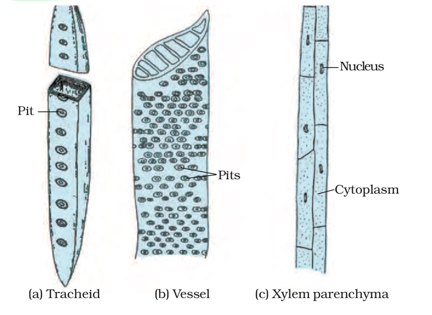
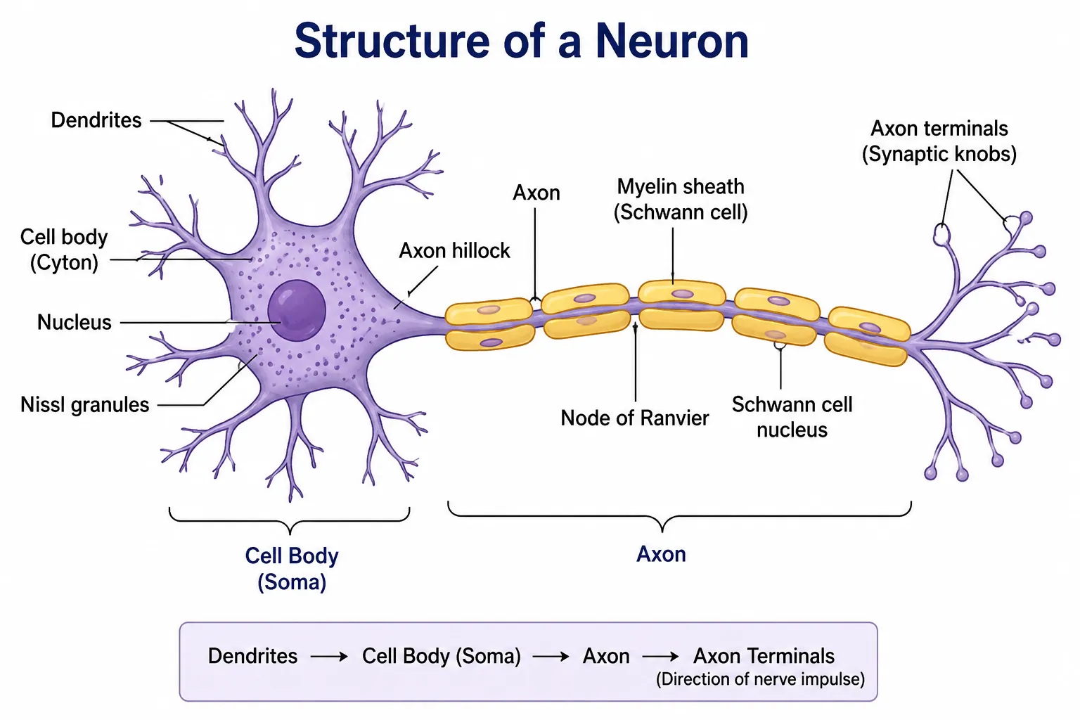
📊 Graph / Figure Graph / Figure
Biological Organisation Cell Tissue Organ System Levels of structural organization in living organisms
✏️ Solution Complete Solution
Step-by-step Solution  ·  5 steps
  1. A multicellular organism contains numerous cells.
  2. Cells with similar structure and common origin become grouped together.
  3. These grouped cells perform a particular function collectively.
  4. Therefore, tissue is defined as a group of similar cells having a common origin that work together to perform a specific function.
  5. Tissue is a level of biological organisation that lies between cells and organs.
🎯 Exam Significance Exam Significance
  • This is one of the most frequently asked one-mark definitions in Class IX examinations.
  • Students should use the keywords "group of similar cells" and "perform a specific function" to obtain full marks.
  • The definition forms the foundation for understanding plant and animal tissues in subsequent questions.
🔑 Key Takeaways Key Takeaways
Key Takeaways  ·  5 points
  1. A tissue is a group of similar cells having a common origin.

  2. The cells in a tissue work together to perform a specific function.

  3. Tissues provide division of labour in multicellular organisms.

  4. Tissue is an organisational level between cells and organs.

  5. Specialisation of tissues increases the efficiency of living organisms.

↑ Top
1 / 15  ·  7%
Q2 →
Q2
NUMERIC3 marks
How many types of elements together make up the xylem tissue? Name them.
📘 Concept & Theory Concept and Theory

Xylem is a complex permanent tissue found in vascular plants. A complex tissue is made up of more than one type of cell that work together to perform a common function.

The primary function of xylem is the conduction of water and dissolved minerals from roots to different parts of the plant. It also provides mechanical strength and support to the plant body.

NCERT states that xylem consists of four different types of elements. These elements work in a coordinated manner to perform transportation and supportive functions.

🗺️ Solution Roadmap Step-by-step Plan
  1. Recall that xylem is a complex permanent tissue.

  2. Determine the number of elements present in xylem.

  3. Name all the elements one by one.

  4. Mention the function of each element briefly.

✏️ Solution Complete Solution
Step-by-step Solution  ·  5 steps
  1. Xylem is a complex permanent tissue because it consists of more than one type of cell.
  2. Xylem is made up of four different elements and not three.
  3. The four elements of xylem are:
  4. Xylem Tissue
    • Tracheids: These are elongated, tapering, dead cells with thick and lignified walls. They conduct water and minerals and also provide mechanical support.
    • Vessels (Xylem Vessels): These are long, tube-like dead cells arranged one above another. They form continuous channels that efficiently transport water and minerals throughout the plant.
    • Xylem Parenchyma: These are the only living cells of xylem. They store food materials such as starch and assist in the lateral conduction of water.
    • Xylem Fibres: These are dead, thick-walled, elongated cells that provide mechanical strength and rigidity to the plant body.
  5. Therefore, xylem tissue is composed of four elements: Tracheids, Vessels, Xylem Parenchyma, and Xylem Fibres.
🎯 Exam Significance Exam Significance
  • This is a frequently asked two-mark question from the chapter Tissues.
  • Students often lose marks by mentioning only three elements and forgetting xylem fibres.
  • Writing the names along with one function of each element helps in obtaining full marks.
  • The question is also important for diagram-based and differentiation questions involving xylem and phloem.
🔑 Key Takeaways Key Takeaways
Key Takeaways  ·  6 points
  1. Xylem is a complex permanent tissue.

  2. Xylem performs conduction of water and minerals and provides mechanical support.

  3. Xylem consists of four elements and not three.

  4. The four xylem elements are Tracheids, Vessels, Xylem Parenchyma, and Xylem Fibres.

  5. Xylem Parenchyma is the only living element of xylem.

  6. Tracheids, Vessels, and Xylem Fibres are dead at maturity.

← Q1
2 / 15  ·  13%
Q3 →
Q3
NUMERIC3 marks
How are simple tissues different from complex tissues in plants?
📘 Concept & Theory Concept and Theory

Plants contain different kinds of permanent tissues that perform specialised functions. Based on the types of cells present, permanent tissues are classified into simple tissues and complex tissues.

Simple tissues consist of only one kind of similar cells performing the same function. In contrast, complex tissues consist of more than one type of cell that work together to perform a common function, mainly transportation of materials in plants.

Complex tissues are often called vascular tissues because they form the conducting system of plants. The two vascular tissues are xylem and phloem.

🗺️ Solution Roadmap Step-by-step Plan
  1. Define simple tissues.

  2. Define complex tissues

  3. Compare them on the basis of cell composition, functions, structure, and examples.

  4. Present the differences in tabular form.

📊 Graph / Figure Graph / Figure
Plant Tissues Simple Tissues vs. Complex Tissues SIMPLE TISSUES Made of one cell type performing one function Parenchyma Thin cell walls, large intercellular spaces Function Storage, photosynthesis (chlorenchyma) Found in: cortex, pith Collenchyma Elongated, corners thickened with pectin/cellulose Function Flexibility and mechanical support to growing parts Found in: stem, leaf stalks Sclerenchyma Thick lignified walls, no protoplasm, dead cells Function Mechanical strength and rigidity to plant parts Found in: husk, seed coat VS COMPLEX TISSUES Made of more than one cell type working together Xylem Conducting (water-carrying) tissue Tracheids Tapering dead cells, water transport in gymnosperms Vessels Tube-like, perforated end walls, main water conduit Xylem fibres Sclerenchymatous, give mechanical strength Xylem parenchyma Living cells, store food Phloem Conducting (food-carrying) tissue Sieve tubes Tube cells with perforated sieve plates, carry food Companion cells Living cells beside sieve tubes, assist conduction Phloem fibres Sclerenchymatous, provide support Phloem parenchyma Stores food, absent in monocots NCERT Biology — Tissues
✏️ Solution Complete Solution
Step-by-step Solution  ·  4 steps
  1. Simple tissues are made up of only one type of similar cells that perform the same function.
  2. Complex tissues are made up of different types of cells that work together to perform a common function.
  3. The differences between simple and complex tissues are as follows:
  4. Difference between Simple and Complex Tissues

    Properties Simple Tissue Complex Tissue
    Definition A tissue made up of only one type of similar cells performing the same function. A tissue made up of different types of cells working together to perform a common function.
    Cell Composition Made up of a single, homogeneous type of cell. Composed of multiple different cell types arranged into a single tissue.
    Nature of Cells Cells are structurally and functionally similar. Cells differ in structure and perform different roles within the same tissue.
    Function Performs basic and uniform functions such as storage, photosynthesis, and mechanical support. Performs specialised functions, mainly the transport of water, minerals, and food throughout the plant.
    Origin and Structure All cells have a common origin and a similar structure. Different cells possess different structures and may originate from different cell types.
    Examples Parenchyma, Collenchyma and Sclerenchyma. Xylem and Phloem.
🎯 Exam Significance Exam Significance
  • This is one of the most important comparison questions from the chapter Tissues.
  • The question is frequently asked as a two-mark or three-mark question in school and board examinations.
  • Students should present the answer in a tabular form because it improves clarity and helps secure full marks.
  • Remember the keywords: homogeneous, heterogeneous, same function, and transportation.
🔑 Key Takeaways Key Takeaways
Key Takeaways  ·  7 points
  1. Simple tissues consist of only one type of similar cells.

  2. Complex tissues consist of different types of cells working together.

  3. Simple tissues mainly perform storage, photosynthesis, and support functions.

  4. Complex tissues mainly perform transportation of water, minerals, and food.

  5. Parenchyma, Collenchyma, and Sclerenchyma are simple tissues.

  6. Xylem and Phloem are complex or vascular tissues.

  7. Complex tissues exhibit division of labour among their different cell types.

← Q2
3 / 15  ·  20%
Q4 →
Q4
NUMERIC3 marks
Differentiate between parenchyma, collenchyma and sclerenchyma based on their cell wall.
📘 Concept & Theory Concept and Theory

Parenchyma, collenchyma, and sclerenchyma are the three types of simple permanent tissues found in plants. Although all three tissues are made up of similar cells performing related functions, they differ mainly in the nature and thickness of their cell walls.

The cell wall provides shape, strength, protection, and support to plant cells. Different substances are deposited in the cell walls of different tissues according to their functions.

  • Parenchyma has thin cell walls because it mainly performs storage, photosynthesis, and packing functions.
  • Collenchyma possesses unevenly thickened walls that provide both support and flexibility to growing plant parts.
  • Sclerenchyma has very thick and lignified walls that provide great mechanical strength and rigidity.
🗺️ Solution Roadmap Step-by-step Plan
  1. Recall that all three are simple permanent tissues.

  2. Identify the composition and thickness of their cell walls.

  3. Compare the tissues on the basis of wall characteristics.

  4. Present the differences in tabular form.

📊 Graph / Figure Graph / Figure
Cell Walls of Simple Permanent Tissues Wall composition determines each tissue's role Parenchyma Thin, uniform wall WALL TYPE Thin and uniformly thick throughout the cell COMPOSITION Made of cellulose only KEY FEATURE Living protoplasm, large vacuole, prominent intercellular spaces WHY THIS WALL? Thin walls allow easy diffusion of gases and water — ideal for storage, photosynthesis, and secretion functions Collenchyma Thickened corners WALL TYPE Unevenly thick — corners are thickened, faces are thin COMPOSITION Cellulose, hemicellulose and pectin deposits KEY FEATURE Living cells, elongated, little or no intercellular space WHY THIS WALL? Corner thickening gives mechanical strength while staying flexible — supports growing stems and leaf stalks without restricting growth Sclerenchyma Thick lignified wall narrow/no lumen WALL TYPE Very thick, uniformly deposited — lumen almost obliterated COMPOSITION Heavily lignified — cellulose + lignin KEY FEATURE Dead cells, no protoplasm, tapering ends WHY THIS WALL? Lignin makes the wall rigid and waterproof — gives hardness and mechanical strength to husks, nutshells, seed coats NCERT Biology — Tissues
Cell Walls of Simple Permanent Tissues
✏️ Solution Complete Solution
Step-by-step Solution  ·  4 steps
  1. Parenchyma cells require flexibility and storage capacity, so their cell walls remain thin and are primarily made of cellulose.
  2. Collenchyma cells provide support to growing parts of plants. Therefore, their cell walls become unevenly thickened, especially at the corners, due to the deposition of cellulose, pectin, and hemicellulose.
  3. Sclerenchyma cells provide mechanical strength and rigidity. Hence, their cell walls become extremely thick and lignified due to the deposition of lignin.
  4. Tissue Nature of Cell Wall
    Parenchyma Thin, primary cell wall made mainly of cellulose.
    Collenchyma Cell wall is unevenly thickened, particularly at the corners, due to deposition of cellulose, pectin, and hemicellulose.
    Sclerenchyma Very thick, rigid, and lignified cell wall due to deposition of lignin.
🎯 Exam Significance Exam Significance
  • This is a frequently asked two-mark or three-mark comparison question from the chapter Tissues.
  • Students should specifically mention the substances deposited in the cell walls: cellulose, pectin, hemicellulose, and lignin.
  • The terms thin, unevenly thickened, and lignified are important keywords and should be included in the answer.
  • This concept is often linked with questions on the functions and locations of simple permanent tissues.
🔑 Key Takeaways Key Takeaways
Key Takeaways  ·  6 points
  1. Parenchyma has thin cell walls made mainly of cellulose.

  2. Collenchyma has unevenly thickened cell walls due to deposition of cellulose, pectin, and hemicellulose.

  3. Sclerenchyma possesses very thick, rigid, and lignified cell walls.

  4. Parenchyma mainly performs storage and metabolic activities.

  5. Collenchyma provides support with flexibility to growing plant parts.

  6. Sclerenchyma provides maximum mechanical strength and rigidity to plants.

← Q3
4 / 15  ·  27%
Q5 →
Q5
NUMERIC3 marks
What are the functions of the stomata?
📘 Concept & Theory Concept and Theory

Stomata are minute pores present mainly on the epidermis of leaves and young green stems. Each stomatal pore is surrounded by two specialised, kidney-shaped cells called guard cells.

The opening and closing of stomata are controlled by the guard cells. When guard cells absorb water, they become turgid and the stomatal pore opens. When they lose water, they become flaccid and the pore closes.

Stomata play an important role in maintaining the physiological activities of plants. They regulate the exchange of gases and the loss of water from the plant body.

🗺️ Solution Roadmap Step-by-step Plan
  1. Define stomata and mention their location.

  2. State that they are surrounded by guard cells.

  3. Explain their role in gaseous exchange.

  4. Explain their role in transpiration.

📊 Graph / Figure Graph / Figure
Structure and Functions of Stomata Tiny pores that let the leaf breathe STRUCTURE OF A STOMATAL UNIT Epidermal cells (surface view) Guard cells bean-shaped, living, thick inner wall Stomatal pore OPEN STOMATA Guard cells turgid — pore opens wide CLOSED STOMATA Guard cells flaccid — pore shuts How Opening & Closing Works Water enters guard cells, making them turgid. The thin outer wall stretches more than the thick inner wall, so the cells bow outward and the pore opens. When guard cells lose water and become flaccid, this bowing reverses and the pore closes. FUNCTIONS OF STOMATA 1 Gas Exchange CO₂ enters for photosynthesis; O₂ released as a by-product 2 Transpiration Water vapour escapes, cooling the plant and pulling water and minerals up from the roots 3 Water Regulation Guard cells open or close the pore to control water loss based on light, humidity, and temperature 4 Respiration Allows continuous O₂ uptake and CO₂ release for cellular respiration, day and night Stomata occur mainly on the lower epidermis of leaves, surrounded by epidermal cells that limit water loss while keeping gas exchange efficient
Functions of Stomata
✏️ Solution Complete Solution
Step-by-step Solution  ·  4 steps
  1. Stomata are tiny pores present on the surface of leaves and young stems.
  2. Each stomatal pore is surrounded by two kidney-shaped guard cells that regulate its opening and closing.
  3. The functions of stomata are as follows:
    • Gaseous Exchange: Stomata facilitate the exchange of gases between the plant and the atmosphere. Carbon dioxide enters the leaf for photosynthesis, while oxygen and carbon dioxide move out during respiration and photosynthesis.
    • Transpiration: Stomata facilitate transpiration, which is the loss of excess water in the form of water vapour from the aerial parts of the plant, especially through the leaves.
  4. Therefore, stomata perform two major functions: gaseous exchange and transpiration.
🌟 Importance of Transpiration
  • Helps in the upward movement of water and minerals from roots to leaves.
  • Maintains the water balance of the plant.
  • Prevents overheating by cooling the plant body.
  • Removes excess water from plant tissues.
🎯 Exam Significance Exam Significance
  • This is a frequently asked two-mark question from the chapter Tissues.
  • Students should mention both functions of stomata to obtain full marks.
  • The keywords gaseous exchange and transpiration should always be included in the answer.
  • The question is often linked with diagrams of stomata and the role of guard cells.
🔑 Key Takeaways Key Takeaways
Key Takeaways  ·  6 points
  1. Stomata are minute pores present mainly on the epidermis of leaves.

  2. Each stomatal pore is surrounded by two kidney-shaped guard cells.

  3. Guard cells regulate the opening and closing of stomata.

  4. Stomata facilitate gaseous exchange between plants and the atmosphere.

  5. Stomata facilitate transpiration, which involves the loss of water as water vapour.

  6. Transpiration helps in cooling the plant and transporting water and minerals.

← Q4
5 / 15  ·  33%
Q6 →
Q6
NUMERIC3 marks
Diagrammatically show the difference between the three types of muscle fibres.
📘 Concept & Theory Concept and Theory

Muscle tissue is a specialised animal tissue responsible for movement and locomotion. The cells of muscular tissue are elongated and are called muscle fibres. These fibres have the ability to contract and relax, thereby producing movement.

Based on their structure, location, and mode of action, muscle fibres are classified into three types:

  1. Striated (Skeletal or Voluntary) Muscles
  2. Smooth (Unstriated or Involuntary) Muscles
  3. Cardiac Muscles

These muscle fibres differ in appearance, presence of striations, number of nuclei, branching pattern, and control of movement.

🗺️ Solution Roadmap Step-by-step Plan
  1. Identify the three types of muscle fibres.

  2. Represent each muscle fibre diagrammatically.

  3. Highlight the distinguishing features of each muscle fibre.

  4. Compare them on the basis of structure and function.

📊 Graph / Figure Graph / Figure
Muscle Tissue
types of muscle fibres
✏️ Solution Complete Solution
Step-by-step Solution  ·  4 steps
  1. Striated muscles are long, cylindrical, unbranched fibres having alternate dark and light bands. They possess many nuclei and are under voluntary control.
  2. Smooth muscles are spindle-shaped fibres with pointed ends. They do not possess striations and contain a single nucleus. Their activities are involuntary.
  3. Cardiac muscles are cylindrical and branched fibres. They possess faint striations and usually have one nucleus. They are involuntary muscles found only in the heart.
  4. Characteristic Striated Muscle Smooth Muscle Cardiac Muscle
    Shape Long, cylindrical and unbranched Spindle-shaped with pointed ends Cylindrical and branched
    Striations Present Absent Present
    Nuclei Many nuclei Single nucleus Usually one nucleus
    Control Voluntary Involuntary Involuntary
    Location Attached to bones Walls of internal organs such as stomach and intestine Heart
🎯 Exam Significance Exam Significance
  • This is an important three-mark and diagram-based question from the chapter Tissues.
  • Students should remember the terms striated, unstriated, branched, voluntary, and involuntary.
  • Labelling the diagrams properly and writing one or two distinguishing features of each muscle fibre helps in securing full marks.
  • The question is frequently linked with the functions and locations of muscle tissues.
🔑 Key Takeaways Key Takeaways
Key Takeaways  ·  6 points
  1. Muscle fibres are specialised cells capable of contraction and relaxation.

  2. There are three types of muscle fibres: Striated, Smooth, and Cardiac.

  3. Striated muscles are voluntary, multinucleate, and possess alternate light and dark bands.

  4. Smooth muscles are spindle-shaped, uninucleate, and involuntary.

  5. Cardiac muscles are branched, striated, involuntary, and found only in the heart.

  6. All muscle tissues work together to produce movement and perform essential life processes.

← Q5
6 / 15  ·  40%
Q7 →
Q7
NUMERIC3 marks
What is the specific function of the cardiac muscle?
📘 Concept & Theory Concept and Theory

Cardiac muscle is a specialised type of muscular tissue found exclusively in the walls of the heart. It is made up of branched, cylindrical, and striated muscle fibres that are interconnected and work in a highly coordinated manner.

Unlike skeletal muscles, cardiac muscles are involuntary muscles. Their contractions are not under conscious control. Cardiac muscle fibres contain contractile proteins that enable them to contract and relax continuously throughout life without fatigue.

Since the heart has to function continuously, cardiac muscles are richly supplied with blood vessels and have abundant mitochondria to meet their high energy requirements.

🗺️ Solution Roadmap Step-by-step Plan
  1. Identify the location of cardiac muscles.

  2. Recall that cardiac muscles are involuntary and rhythmic.

  3. State their specific role in the functioning of the heart.

  4. Explain how their contraction and relaxation help in blood circulation.

📊 Graph / Figure Graph / Figure
Specific Function of Cardiac Muscle Blood Pumped to the Body Rhythmic Contraction Involuntary Heartbeat
✏️ Solution Complete Solution
Step-by-step Solution  ·  6 steps
  1. Cardiac muscles are present only in the walls of the heart.
  2. These muscle fibres undergo rhythmic and involuntary contraction and relaxation.
  3. The rhythmic contractions of cardiac muscles generate the heartbeat.
  4. The continuous contraction and relaxation of cardiac muscles pump blood from the heart to different parts of the body and return blood back to the heart.
  5. Therefore, the specific function of cardiac muscle is to bring about the rhythmic, involuntary contractions of the heart that continuously pump blood throughout the body.
  6. Why is this Function Important?
    • Ensures a continuous supply of oxygen and nutrients to body cells.
    • Helps transport hormones and other essential substances throughout the body.
    • Facilitates the removal of carbon dioxide and metabolic wastes.
    • Maintains proper circulation and functioning of all organs.
🎯 Exam Significance Exam Significance
  • This is an important one-mark or two-mark conceptual question from the chapter Tissues.
  • Students should specifically mention the terms rhythmic, involuntary contraction, and pumping of blood.
  • Writing only "causes movement" is incomplete because the question asks for the specific function of cardiac muscle.
  • This concept is frequently linked with questions on the characteristics of cardiac muscles and their comparison with skeletal and smooth muscles.
🔑 Key Takeaways Key Takeaways
Key Takeaways  ·  6 points
  1. Cardiac muscles are found only in the walls of the heart.

  2. They are involuntary and work continuously throughout life.

  3. They contract and relax rhythmically to produce the heartbeat.

  4. Their specific function is to pump blood continuously throughout the body.

  5. Cardiac muscles are richly supplied with blood vessels and contain numerous mitochondria.

  6. The continuous pumping action of cardiac muscles sustains life by maintaining blood circulation.

← Q6
7 / 15  ·  47%
Q8 →
Q8
NUMERIC3 marks
Differentiate between striated, unstriated and cardiac muscles based on their structure and site/location in the body.
📘 Concept & Theory Concept and Theory

Muscular tissue is a specialised animal tissue composed of elongated cells called muscle fibres. These fibres contain contractile proteins that enable them to contract and relax, thereby producing movement.

Based on their structure and function, muscle fibres are classified into three types: striated muscles, unstriated muscles, and cardiac muscles.

These muscles differ in their appearance, branching pattern, number of nuclei, presence or absence of striations, and the parts of the body where they are found.

🗺️ Solution Roadmap Step-by-step Plan
  1. Recall the three types of muscle tissues.

  2. Compare their structural features.

  3. Compare the sites where each muscle tissue is found.

  4. Present the differences in a tabular form.

📊 Graph / Figure Graph / Figure
Structure and Location of Muscle Fibres Three types of muscle tissue in the body SKELETAL (STRIATED) MUSCLE STRUCTURE Long, cylindrical, unbranched fibres NUCLEUS Many, pushed to the periphery PATTERN Striated (alternating light/dark bands) CONTROL Voluntary — consciously controlled LOCATION Attached to bones — limbs, face, body wall FUNCTION Body movement & posture SMOOTH (UNSTRIATED) MUSCLE STRUCTURE Spindle-shaped, tapering at both ends NUCLEUS Single, oval, centrally located PATTERN Unstriated — no visible banding CONTROL Involuntary — not under conscious control LOCATION Walls of stomach, intestine, blood vessels, ureters FUNCTION Slow, sustained movement CARDIAC MUSCLE STRUCTURE Branched, cylindrical, joined end to end NUCLEUS Single, centrally located PATTERN Striated, with intercalated discs CONTROL Involuntary — rhythmic, never tires LOCATION Found only in the walls of the heart FUNCTION Pumps blood throughout life NCERT Biology — Tissues
✏️ Solution Complete Solution
Step-by-step Solution  ·  4 steps
  1. Striated muscles are long, cylindrical, unbranched, and multinucleate fibres showing alternate light and dark bands called striations.
  2. Unstriated muscles are long, spindle-shaped, unbranched fibres that lack striations and possess a single nucleus.
  3. Cardiac muscles are cylindrical and branched fibres that form a network. They possess faint striations, usually a single nucleus, and are connected by specialised junctions called intercalated discs.
  4. Difference between Striated, Unstriated and Cardiac Muscles

    Properties Striated Muscles Unstriated Muscles Cardiac Muscles
    Structure Long, cylindrical and unbranched fibres. They exhibit alternate light and dark bands called striations and are multinucleate. Long, spindle-shaped and unbranched fibres. They do not exhibit striations and are usually uninucleate. Cylindrical and branched fibres that form a network. They possess faint striations, are usually uninucleate, and are connected by intercalated discs.
    Site/Location Found attached to bones and present in the limbs, face, tongue, and other body parts involved in voluntary movements. Found in the walls of internal organs such as the stomach, intestine, urinary bladder, uterus, and blood vessels. Found exclusively in the walls of the heart.
🎯 Exam Significance Exam Significance
  • This is one of the most important three-mark comparison questions from the chapter Tissues.
  • Students should write the answer in tabular form to improve presentation and score full marks.
  • The keywords striations, branched, multinucleate, uninucleate, and intercalated discs should be included in the answer.
  • The question is frequently linked with questions on voluntary and involuntary movements.
🔑 Key Takeaways Key Takeaways
Key Takeaways  ·  6 points
  1. There are three types of muscle tissues: Striated, Unstriated, and Cardiac muscles.

  2. Striated muscles are cylindrical, unbranched, multinucleate, and attached to bones.

  3. Unstriated muscles are spindle-shaped, unbranched, uninucleate, and present in the walls of internal organs.

  4. Cardiac muscles are branched, faintly striated, usually uninucleate, and found only in the heart.

  5. Intercalated discs are characteristic features of cardiac muscles.

  6. The structure of each muscle tissue is closely related to its specific function.

← Q7
8 / 15  ·  53%
Q9 →
Q9
NUMERIC3 marks
Draw a labelled diagram of a neuron.
📊 Graph / Figure Graph / Figure
Structure of Neuron
← Q8
9 / 15  ·  60%
Q10 →
Q10
NUMERIC2 marks
Name the following.

(a) Tissue that forms the inner lining of our mouth. Squamous epithelium Tissue
(b) Tissue that connects muscle to bone in humans. Tendons Tissue
(c) Tissue that transports food in plants. Phloem Tissue
(d) Tissue that stores fat in our body. Adipose Tissue
(e) Connective tissue with a fluid matrix. Blood Tissue
(f) Tissue present in the brain. Nervous Tissue
📘 Concept & Theory Concept and Theory

Different tissues in plants and animals are specialised to perform particular functions. The structure and organisation of each tissue enable it to perform its function efficiently. Therefore, by understanding the function mentioned in the question, we can identify the correct tissue.

This question tests the concept of division of labour in multicellular organisms, where different tissues are specialised for different activities such as protection, support, transport, storage, and coordination.

🗺️ Solution Roadmap Step-by-step Plan
  1. Identify the function mentioned in each statement.

  2. Recall the tissue specialised to perform that function.

  3. Name the correct tissue.

  4. Mention one important characteristic or function of the tissue.

✏️ Solution Complete Solution
Step-by-step Solution  ·  7 steps
  1. (a) Tissue that forms the inner lining of our mouth
  2. The inner lining of the mouth requires a protective covering against friction and wear and tear. Therefore, it is lined by Squamous Epithelium (Squamous Epithelial Tissue).
  3. (b) Tissue that connects muscle to bone in humans
  4. Muscles need to be firmly attached to bones so that movement can occur. This function is performed by strong and flexible connective tissue called Tendon.
  5. (c) Tissue that transports food in plants
  6. Plants transport prepared food from leaves to other parts through the conducting tissue called Phloem.
  7. (d) Tissue that stores fat in our body
  8. Fat is stored in specialised connective tissue composed of fat-storing cells called adipocytes. This tissue is known as Adipose Tissue.
  9. (e) Connective tissue with a fluid matrix
  10. Blood is a connective tissue because it connects different parts of the body by transporting substances. Its matrix, called plasma, is fluid in nature.
  11. (f) Tissue present in the brain
  12. The brain is mainly composed of specialised cells called neurons that receive and transmit nerve impulses. Therefore, the tissue present in the brain is Nervous Tissue.
  13. Summary Table
  14. Question Answer Main Function
    (a) Inner lining of the mouth Squamous Epithelium Protection against friction and wear
    (b) Connects muscle to bone Tendon Attachment and transmission of muscular force
    (c) Transports food in plants Phloem Transport of prepared food
    (d) Stores fat Adipose Tissue Storage of fat and insulation
    (e) Connective tissue with fluid matrix Blood Transportation of materials
    (f) Tissue present in the brain Nervous Tissue Transmission of nerve impulses
🎯 Exam Significance Exam Significance
  • This is an important one-mark and objective-type question from the chapter Tissues.
  • The question tests students' understanding of the functions of various tissues.
  • Students should remember the association between a tissue and its specialised function.
  • Such questions are frequently asked in fill-in-the-blanks, matching, and MCQ formats.
🔑 Key Takeaways Key Takeaways
Key Takeaways  ·  7 points
  1. Squamous epithelium forms protective linings of body organs.

  2. Tendons connect muscles to bones.

  3. Phloem transports prepared food in plants.

  4. Adipose tissue stores fat and acts as an insulator.

  5. Blood is a connective tissue having a fluid matrix called plasma.

  6. Nervous tissue consists of neurons that conduct nerve impulses.

  7. Different tissues are specialised for different functions, demonstrating division of labour in multicellular organisms.

← Q9
10 / 15  ·  67%
Q11 →
Q11
NUMERIC3 marks
Identify the type of tissue in the following: skin, bark of a tree, bone, lining of kidney tubule, vascular bundle.
📘 Concept & Theory Concept and Theory

Different tissues in plants and animals are specialised to perform specific functions. The structure, arrangement, and location of tissues enable them to carry out their functions efficiently.

To identify a tissue correctly, we should first understand the function performed by the organ or body part in which it is present. Protective organs contain protective tissues, supporting organs contain supportive tissues, and conducting structures contain transport tissues.

This question is based on the concept of division of labour in multicellular organisms, where different tissues perform different specialised functions.

🗺️ Solution Roadmap Step-by-step Plan
  1. Identify the structure or organ mentioned in the question.

  2. Recall its primary function.

  3. Determine the tissue best suited to perform that function.

  4. Name the tissue and mention its role briefly.

✏️ Solution Complete Solution
Step-by-step Solution  ·  6 steps
  1. (a) Skin
  2. The skin forms the outer protective covering of the body. It protects the body from injury, dehydration, and infection. Therefore, the tissue present in the skin is Epithelial Tissue.
  3. (b) Bark of a Tree
  4. The bark acts as a protective covering of the stem and prevents water loss and mechanical injury. The protective tissue present in the bark is called Cork (Phellem).
  5. (c) Bone
  6. Bones provide support, shape, and protection to the body and also serve as attachment sites for muscles. Therefore, bones are composed of Connective Tissue.
  7. (d) Lining of Kidney Tubules
  8. Kidney tubules perform absorption and secretion of substances. Such functions require cube-shaped cells. Therefore, the lining of kidney tubules consists of Cuboidal Epithelium (Cuboidal Epithelial Tissue).
  9. (e) Vascular Bundle
  10. Vascular bundles consist of xylem and phloem tissues that transport water, minerals, and food throughout the plant. Since they are composed of different types of cells working together, they belong to Complex Permanent Tissue.
  11. Summary Table
  12. Structure Type of Tissue Main Function
    Skin Epithelial Tissue Protection of the body surface
    Bark of a Tree Cork (Phellem) Protection and prevention of water loss
    Bone Connective Tissue Support, protection, and attachment of muscles
    Lining of Kidney Tubules Cuboidal Epithelium Absorption and secretion
    Vascular Bundle Complex Permanent Tissue Transport of water, minerals, and food
🎯 Exam Significance Exam Significance
  • This is an important one-mark and objective-type question from the chapter Tissues.
  • The question tests the understanding of the location and functions of various tissues.
  • Students should remember the relationship between tissue structure and its function.
  • Similar questions frequently appear in fill-in-the-blanks, matching, assertion-reasoning, and MCQ formats.
🔑 Key Takeaways Key Takeaways
Key Takeaways  ·  7 points
  1. Skin is composed of protective epithelial tissue.

  2. Bark of a tree contains cork or phellem, which is a protective tissue.

  3. Bone is a supporting connective tissue.

  4. Kidney tubules are lined by cuboidal epithelium specialised for absorption and secretion.

  5. Vascular bundles are complex permanent tissues consisting of xylem and phloem.

  6. The location and function of a tissue are closely related to its structure.

  7. Specialisation of tissues enables efficient functioning of multicellular organisms.

← Q10
11 / 15  ·  73%
Q12 →
Q12
NUMERIC3 marks
Name the regions in which parenchyma tissue is present.
📘 Concept & Theory Concept and Theory

Parenchyma is the most common and widely distributed simple permanent tissue in plants. It consists of living, thin-walled cells with intercellular spaces. Since parenchyma performs functions such as storage, photosynthesis, secretion, and providing support, it is found in many soft parts of the plant body.

🗺️ Solution Roadmap Step-by-step Plan
  1. Recall the nature of parenchyma tissue.

  2. Identify the plant parts that require storage and metabolic activities.

  3. Name the regions where parenchyma is commonly present.

📊 Graph / Figure Graph / Figure
Regions Where Parenchyma Tissue is Present Parenchyma Cortex Pith Leaf Mesophyll Fruits and Seeds
✏️ Solution Complete Solution
Step-by-step Solution  ·  4 steps
  1. Parenchyma is made up of living cells that perform storage and metabolic functions.
  2. Therefore, it is mainly found in the soft regions of plants.
  3. The regions where parenchyma tissue is present are:
    • The cortex of roots and stems.
    • The pith of stems and roots.
    • The mesophyll tissue of leaves.
    • The fleshy parts of fruits.
    • The storage tissues of seeds.
    • The soft tissues present in various plant organs.
  4. Therefore, parenchyma tissue is present mainly in the cortex, pith, mesophyll of leaves, fruits, seeds, and other soft parts of plants.
🎯 Exam Significance Exam Significance
  • This is an important one-mark and two-mark question from the chapter Tissues.
  • Students should remember that parenchyma is the most abundant and widely distributed plant tissue.
  • The terms cortex, pith, and mesophyll are important keywords and should be included in the answer.
  • The question is often linked with the functions and modifications of parenchyma tissue.
🔑 Key Takeaways Key Takeaways
Key Takeaways  ·  6 points
  1. Parenchyma is a living simple permanent tissue.

  2. It is the most abundant and widely distributed tissue in plants.

  3. It is mainly found in the cortex and pith of stems and roots.

  4. It is present in the mesophyll tissue of leaves.

  5. It also occurs in fruits, seeds, and other soft parts of plants.

  6. Its widespread distribution is related to its roles in storage, photosynthesis, secretion, and support.

← Q11
12 / 15  ·  80%
Q13 →
Q13
NUMERIC3 marks
What is the role of epidermis in plants?
📘 Concept & Theory Concept and Theory

The epidermis is the outermost protective layer of cells covering the entire body of a plant. It usually consists of a single layer of tightly packed cells without intercellular spaces. Since it forms the first line of contact between the plant and its surroundings, the epidermis performs several protective and regulatory functions.

The epidermis is specially adapted according to the habitat of the plant. In plants growing in dry habitats, the epidermis is thicker and is covered by a waxy layer called the cuticle, which reduces water loss and protects the plant from harsh environmental conditions.

🗺️ Solution Roadmap Step-by-step Plan
  1. Define the epidermis.

  2. Mention that it forms the outermost protective covering of the plant.

  3. Explain its protective and water-conserving functions.

  4. Mention additional roles such as gaseous exchange and absorption in roots.

📊 Graph / Figure Graph / Figure
PLANT PHYSIOLOGY Roles of Epidermis in Plants CUTICLE (WAXY PROTECTIVE LAYER) Epidermis Protective Outermost Layer of the Plant Body Protection Barrier against injury, pathogens, and pests Water Conservation Waxy cuticle minimizes transpiration / water loss Specialized Cell Adaptations Stomata regulates gas exchange & transpiration Root hairs maximize water & mineral absorption
Roles of Epidermis in Plants
✏️ Solution Complete Solution
Step-by-step Solution  ·  4 steps
  1. The epidermis is a single layer of closely packed cells that forms the outermost covering of the plant.
  2. The cells of the epidermis are usually flat and continuous, with no intercellular spaces, thereby providing protection to the plant body.
  3. The epidermis performs the following important functions:
    • Protection: It protects the entire plant body from mechanical injury, pathogens, and harmful environmental conditions.
    • Prevention of Water Loss: In aerial parts of the plant, epidermal cells secrete a waxy, water-resistant layer called the cuticle. This layer reduces the loss of water through evaporation.
    • Protection in Desert Plants: In xerophytic plants (plants living in dry habitats), the epidermis and cuticle are thicker to minimise water loss and conserve water.
    • Protection Against Pathogens: The cuticle protects the plant from invasion by parasitic fungi and disease-causing microorganisms.
    • Gaseous Exchange: The epidermis bears stomata, which facilitate the exchange of gases and transpiration.
    • Absorption of Water: In roots, epidermal cells form root hairs that absorb water and dissolved minerals from the soil.
  4. Therefore, the epidermis acts as a protective, water-conserving, and regulatory tissue in plants.
🌟 Importance of Cuticle
  • Reduces excessive loss of water.
  • Protects the plant from high temperatures and drying conditions.
  • Provides protection against mechanical injury.
  • Prevents the entry of harmful microorganisms and parasites.
🎯 Exam Significance Exam Significance
  • This is a frequently asked three-mark question from the chapter Tissues.
  • Students should mention the keywords protection, cuticle, prevention of water loss, stomata, and root hairs.
  • Writing the functions in points improves presentation and helps in obtaining full marks.
  • The question is often linked with diagrams of epidermal tissue and stomata.
🔑 Key Takeaways Key Takeaways
Key Takeaways  ·  8 points
  1. The epidermis is the outermost protective layer of the plant body.

  2. It usually consists of a single layer of closely packed cells.

  3. It protects the plant from mechanical injury and pathogens.

  4. The cuticle secreted by epidermal cells reduces water loss.

  5. Desert plants possess a thick epidermis and cuticle to conserve water.

  6. Stomata present in the epidermis facilitate gaseous exchange and transpiration.

  7. Root hairs formed by epidermal cells absorb water and minerals from the soil.

  8. The epidermis acts as a protective, regulatory, and water-conserving tissue.

← Q12
13 / 15  ·  87%
Q14 →
Q14
NUMERIC3 marks
How does the cork act as a protective tissue?
📘 Concept & Theory Concept and Theory

In older stems and roots of plants, the outer epidermis is gradually replaced by another protective tissue called cork or phellem. Cork is produced by a strip of secondary meristematic tissue called the cork cambium (phellogen).

Cork cells undergo several modifications that make them highly suitable for protection. These cells become dead at maturity and are closely packed without intercellular spaces. Their walls become impregnated with a waxy substance called suberin, which makes them impermeable to water and gases.

🗺️ Solution Roadmap Step-by-step Plan
  1. Define cork and mention its origin.

  2. Describe the structural characteristics of cork cells.

  3. Explain the role of suberin.

  4. Relate these features to its protective functions.

📊 Graph / Figure Graph / Figure
Cork as a Protective Tissue Cork (Phellem) • Dead and Compactly Arranged Cells • Cell Walls Contain Suberin • Impermeable to Water and Gases Prevents Water Loss Protection Against Injury Barrier Against Pathogens and Harsh Conditions
✏️ Solution Complete Solution
Step-by-step Solution  ·  6 steps
  1. Cork is a protective tissue formed by the activity of the cork cambium (phellogen), which is a secondary meristem.
  2. Cork consists of dead cells that are compactly arranged and do not possess intercellular spaces.
  3. The walls of cork cells contain a waxy substance called suberin.
  4. Suberin makes the cork cells impermeable to water and gases.
  5. Therefore, cork acts as a protective tissue in the following ways:
    • It prevents excessive loss of water from the plant body.
    • It protects the plant against mechanical injuries.
    • It acts as a barrier against the entry of disease-causing microorganisms and parasites.
    • It protects the inner tissues from extreme environmental conditions.
    • It reduces unnecessary exchange of gases and helps conserve water in older stems and roots.
  6. Hence, cork functions as an efficient protective covering because its dead, compactly arranged, suberised cells form a tough and water-resistant barrier around the plant.
🌟 Why is Suberin Important?
  • It makes cork cells waterproof.
  • It reduces water loss by evaporation.
  • It prevents the entry of harmful microorganisms.
  • It increases the durability and protective nature of cork tissue.
← Q13
14 / 15  ·  93%
Q15 →
Q15
NUMERIC3 marks
Complete the following chart.
📘 Concept & Theory Concept and Theory

Permanent tissues are formed from meristematic tissues after the cells lose their ability to divide and become specialised to perform specific functions. Based on the types of cells present, permanent tissues are classified into simple permanent tissues and complex permanent tissues.

Simple permanent tissues are composed of only one type of similar cells, whereas complex permanent tissues are composed of different kinds of cells working together to perform a common function, mainly transportation.

🗺️ Solution Roadmap Step-by-step Plan
  1. Identify the two major categories of permanent tissues.

  2. Recall the three types of simple permanent tissues.

  3. Recall the two types of complex permanent tissues.

  4. Fill the empty boxes accordingly.

📊 Graph / Figure Graph / Figure
Classification of Permanent Tissues Permanent Tissue Simple Complex Parenchyma Collenchyma Sclerenchyma Xylem Phloem
Completed Chart
✏️ Solution Complete Solution
Step-by-step Solution  ·  4 steps
  1. Permanent tissues are divided into two groups:
    • Simple Permanent Tissues
    • Complex Permanent Tissues
  2. The chart already shows Collenchyma under simple tissues and Xylem under complex tissues.
  3. The remaining types are:
    • Under Simple Permanent Tissues: Parenchyma and Sclerenchyma
    • Under Complex Permanent Tissues: Phloem
  4. Therefore, the completed chart is:
                            Permanent Tissue
                                    │
                                    ├── Simple Permanent Tissue
                                    │       ├── Parenchyma
                                    │       ├── Collenchyma
                                    │       └── Sclerenchyma
                                    │
                                    └── Complex Permanent Tissue
                                            ├── Xylem
                                            └── Phloem
                        
🎯 Exam Significance Exam Significance
  • This is a frequently asked diagram-based and concept-based question from the chapter Tissues.
  • Students should remember the classification of permanent tissues and their examples.
  • Flowcharts and diagrams are often asked in objective and short-answer questions.
  • The keywords Parenchyma, Collenchyma, Sclerenchyma, Xylem, and Phloem should be memorised.
🔑 Key Takeaways Key Takeaways
Key Takeaways  ·  6 points
  1. Permanent tissues are derived from meristematic tissues.

  2. Permanent tissues are classified into simple and complex permanent tissues.

  3. Simple permanent tissues are composed of one type of similar cells

  4. Parenchyma, Collenchyma, and Sclerenchyma are simple permanent tissues.

  5. Complex permanent tissues consist of different types of cells working together.

  6. Xylem and Phloem are complex permanent tissues responsible for transportation in plants.

← Q14
15 / 15  ·  100%
↑ Back to top
🎓

Chapter Complete!

All 15 solutions for Tissues covered.

↑ Review from the top
📚
ACADEMIA AETERNUM तमसो मा ज्योतिर्गमय · Est. 2025
Sharing this chapter
Tissues | Science Class 9 | Academia Aeternum
Tissues | Science Class 9 | Academia Aeternum — Complete Notes & Solutions · academia-aeternum.com
Get complete Class 9 Science Tissues NCERT solutions with detailed explanations, solved examples, images, and tabulated notes. This post provides step-by-step solutions to all NCERT Class 9 Science Tissues textbook exercises, making it easier for students who face difficulty in solving them. To build a stronger foundation, students can also check the links to Tissues Notes and Class 9 Science Tissues MCQs provided at the bottom of the post for quick revision and exam preparation.
🎓 Class 9 📐 Science 📖 NCERT ✅ Free Access 🏆 CBSE · JEE
Share on
academia-aeternum.com/class-9/science/tissues/exercises/ Copy link
💡
Exam tip: Sharing chapter notes with your study group creates a reinforcement loop. Teaching a concept is the fastest path to mastering it.

Recent posts

    Tissues — Learning Resources

    📄 Detailed Notes
    🧠 Practice MCQs
    ✔️ True / False

    Frequently Asked Questions

    Bone and cartilage.

    Areolar tissue joins different tissues and fills spaces between organs.

    Adipose tissue stores fat and provides insulation.

    Blood is a fluid connective tissue that transports oxygen, nutrients, and hormones.

    Plasma, red blood cells, white blood cells, and platelets.

    Bone is a hard connective tissue that provides shape, support, and protection to body.

    Cartilage is a flexible connective tissue present in ear, nose, and joints.

    Ligament connects bone to bone and provides flexibility and strength.

    Tendon connects muscle to bone and helps in movement.

    Muscular tissues help in movement by contraction and relaxation.

    Dendrites, cell body (cyton), and axon.

    Voluntary muscles act under conscious control; involuntary muscles act without conscious effort.

    Get in Touch

    Let's Connect

    Questions, feedback, or suggestions?
    We'd love to hear from you.